Introduction
Immunofluorescence (IF) is essential for students to utilize in basic science research. It is a critical immunochemical technique that allows for the visualization and detection of numerous components in any given tissue or cell type. IF uses specific antibodies tagged with fluorophores, or fluorescent chemical compounds, and can be performed on cultured cells, cell suspensions, or specific targets ranging from tissue samples to entire organisms.
Methods
IF staining begins with fixation, a method that serves to immobilize target antigens and allows antibodies to bind to targeted cellular components while preserving cellular structure. Fixation is necessary to prevent self-digestion and putrefaction. Based on the given antigen and sample type, optimal fixatives can vary, including cross-linking reagents and organic solvents. Cross-linking reagents, such as formaldehyde, bind to cellular and tissue components and form intermolecular and intramolecular methylene cross-links. Organic solvents, like methanol and acetone, remove lipids and dehydrate cells, which can change the natural structure of the sample.
Tissues are embedded in thin sections of paraffin on glass slides in order to solidify the sample for sectioning. This allows dyes, probes, and antibodies to reach the target sites without being blocked by multiple cell layers. Then, the glass slides must be deparaffinized with xylene, followed by ethanol and distilled (DI) water washes for rehydration.
Two methods for antigen retrieval are necessary to restore epitope-antibody reactivity: Protease-Induced Epitope Retrieval (PIER), which uses enzymes, and Heat-Induced Epitope Retrieval (HIER), which uses heat and pressure. The HIER method achieves much higher rates of restoring immunoreactivity, blocking non-target reactive sites to which secondary antibodies would otherwise bind. The goal of antigen retrieval is to unmask the target epitopes and restore antigenicity.
Before the application of antibodies, blocking must be performed on tissue samples to prevent antibodies from binding to non-target epitopes. Blocking buffers include protein solutions, normal serums, and protein-free commercial buffers. Blocking reagents are ideally not affiliated with target epitopes, have high binding rates to non-target reactive sites, and stabilize cellular structure. The solutions bind all proteins present in the sample, causing the antibodies to compete with the blocking protein for target epitopes. This reduces the likelihood of non-specific binding.
Following blocking is the application of antibodies to the tissue sample. Direct (Primary) IF is a staining method where the fluorophore label is conjugated directly to the primary antibody that will bind and react with the target epitope. Although Direct IF is quicker, Indirect (Secondary) IF is utilized widely for its high sensitivity, signal amplification, and ability to detect several target epitopes in the same sample. During Indirect IF, the primary antibody binds to the target epitope, followed by a secondary fluorophore-tagged antibody that recognizes and binds to the primary antibody.
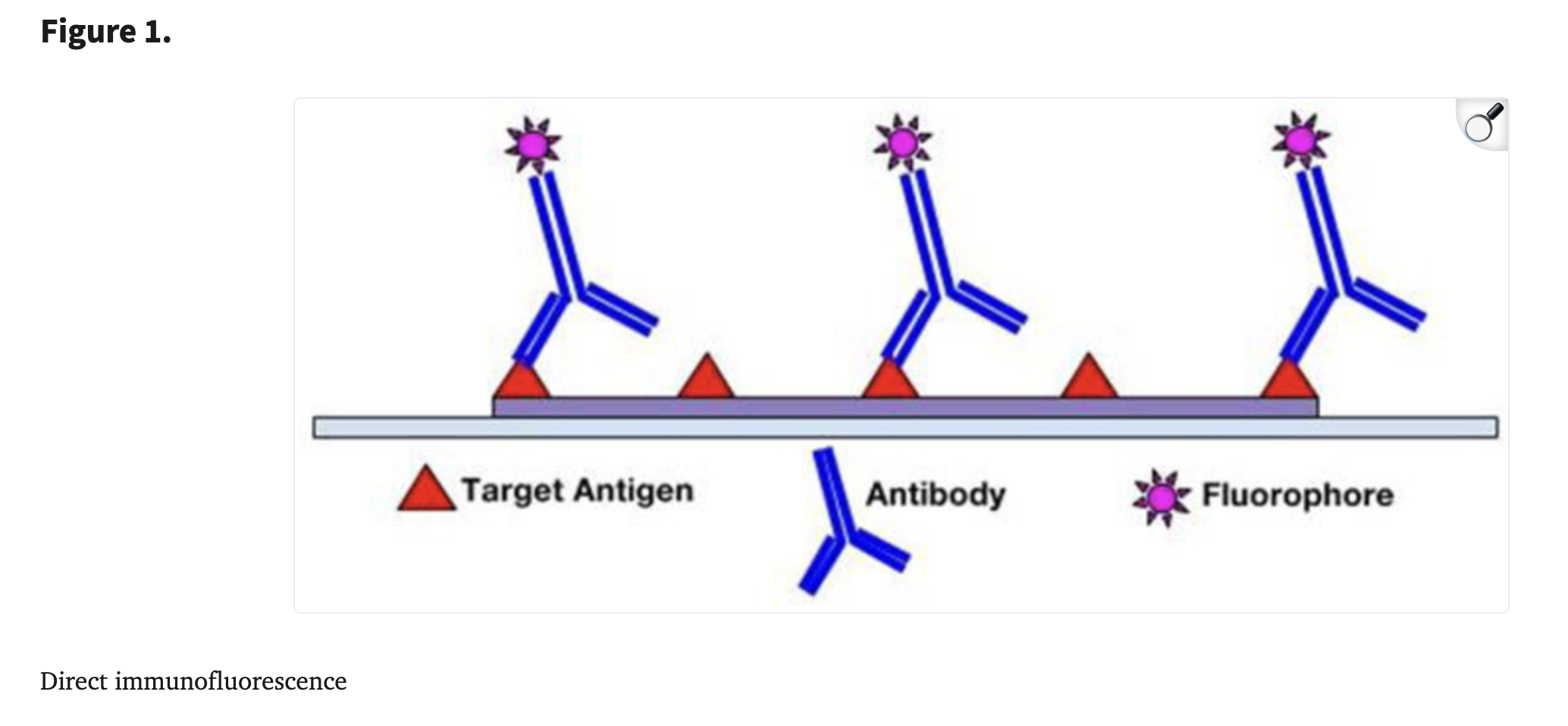
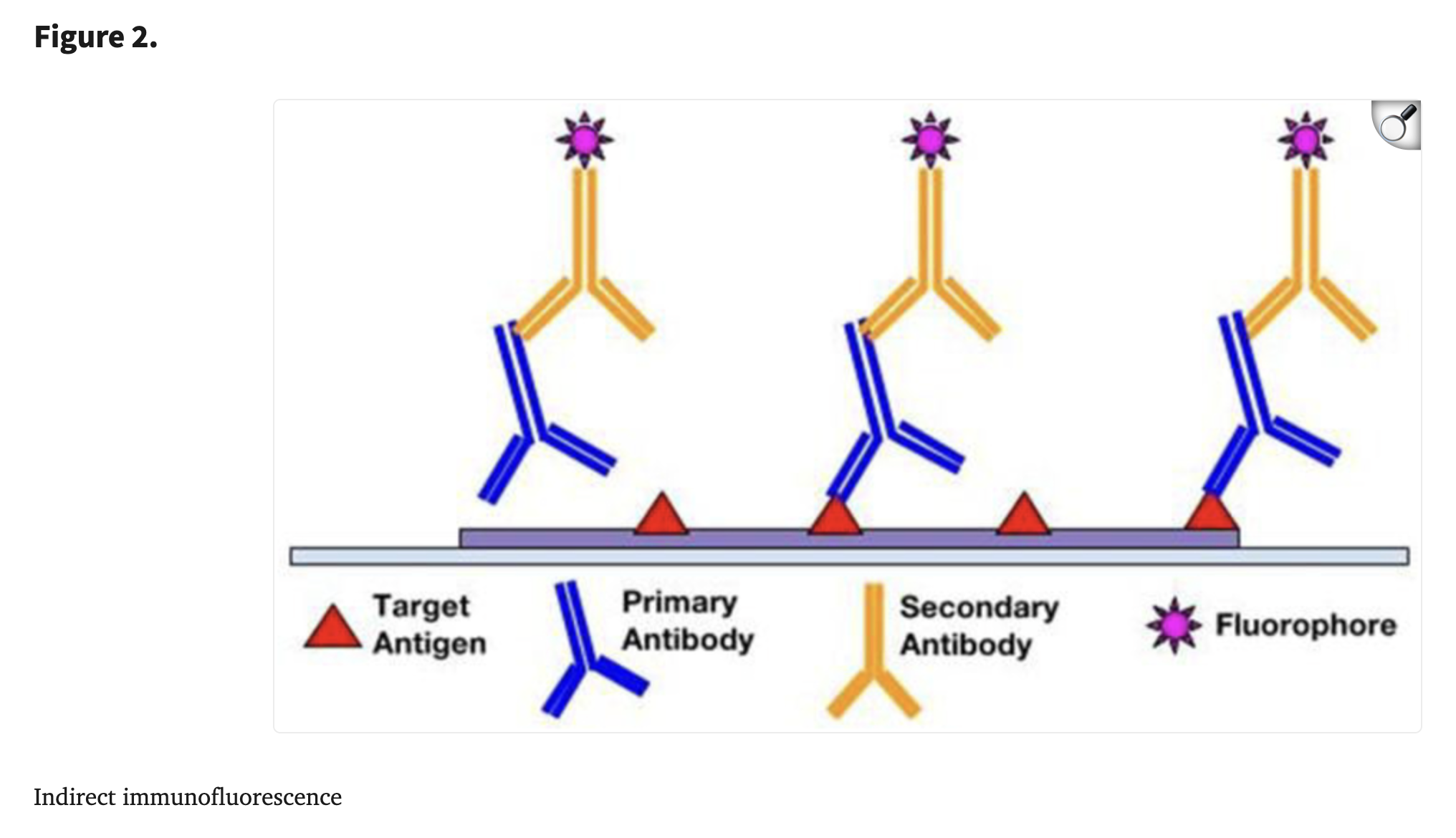
The secondary antibodies pair with fluorescent labels that emit upon photoexcitation, making it easier to identify specific components in the sample. The most commonly used fluorophores are fluorescein isothiocyanate (FITC) and tetramethylrhodamine isothiocyanate (TRITC). The ideal fluorophore is determined by how effectively the epifluorescent microscopes can detect the signal of fluorophores. To minimize photobleaching, photostable fluorophores can be selected, excitation duration and intensity can be reduced, and antifade mounting reagents can be applied.
Conclusion
IF offers many advantages in signal amplification, targeting specificity, and analytical capabilities. It allows the possibility of multiplexing, simultaneously detecting multiple target antigens within the same tissue sample. The process of IF staining is helpful to understand and learn for future research and lab purposes.